39 light microscope with labels
Microscope Labeling Game - Purpose Games About this Quiz. This is an online quiz called Microscope Labeling Game. There is a printable worksheet available for download here so you can take the quiz with pen and paper. This quiz has tags. Click on the tags below to find other quizzes on the same subject. Science. Compound Microscope- Definition, Labeled Diagram, Principle, Parts, Uses The optical microscope often referred to as the light microscope, is a type of microscope that uses visible light and a system of lenses to magnify images of small subjects. The term "compound" in compound microscopes refers to the microscope having more than one lens. Devised with a system of combination of lenses, a compound microscope ...
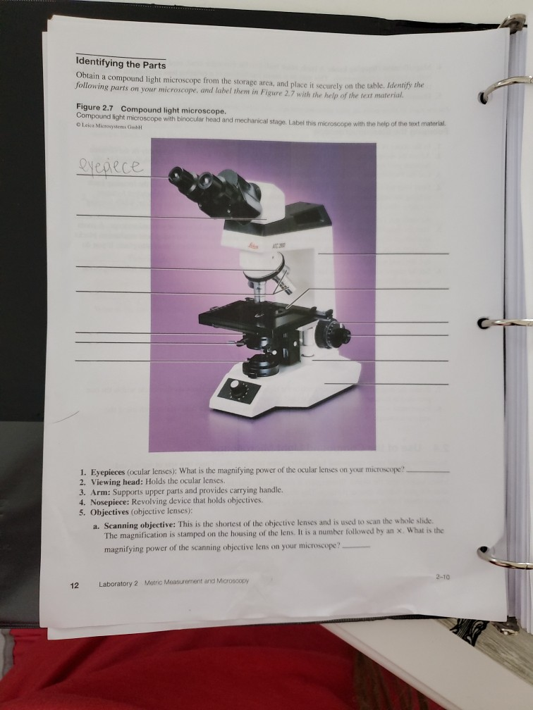
Compound Microscope - Diagram (Parts labelled), Principle and Uses Also called as binocular microscope or compound light microscope, it is a remarkable magnification tool that employs a combination of lenses to magnify the image of a sample that is not visible to the naked eye. Compound microscopes find most use in cases where the magnification required is of the higher order (40 - 1000x).

Light microscope with labels
Microscope Labeling - The Biology Corner 1) Start with scanning (the shortest objective) and only use the COARSE knob . Once it is focused… 2) Switch to low power (medium) and only use the COARSE knob . You may need to recenter your slide. Once it is focused.. 3) Switch to high power (long objective). Microscope Label Worksheets - K12 Workbook Displaying all worksheets related to - Microscope Label. Worksheets are The microscope parts and use, Parts of the light microscope, Label parts of the microscope, Labeling scientific tools microscope name, Parts of the microscope quiz, Use the word list to help you label the 12, Label parts of the microscope answers, Microscope lab. Light Microscope Parts Labeled - 18 images - parts of the microscope ... [Light Microscope Parts Labeled] - 18 images - optical microscopy and specimen using the transmission, microscope with labels clip art at vector clip, solved microscope parts labeling 9 label the image of a c, ,
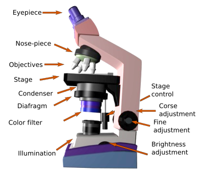
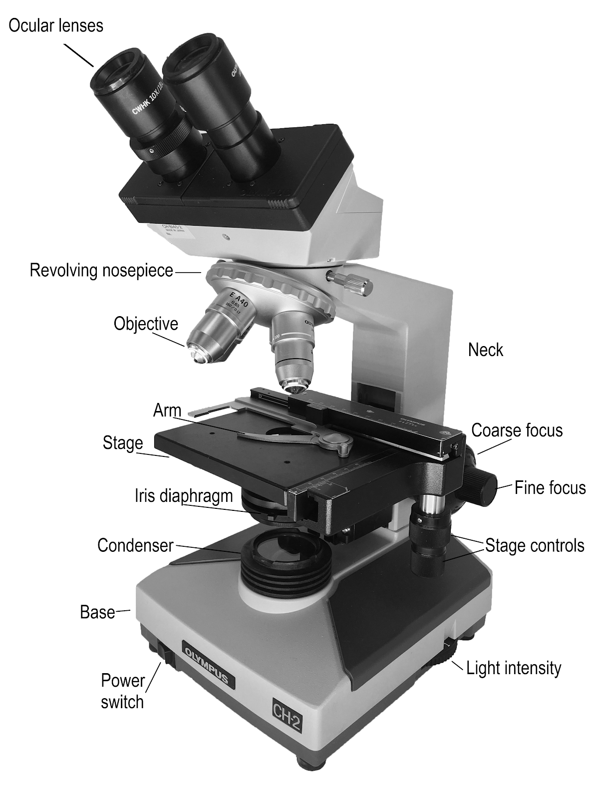
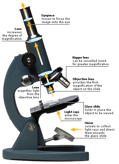
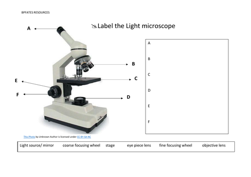
Light microscope with labels. Parts of the Microscope with Labeling (also Free Printouts) Parts of the Microscope with Labeling (also Free Printouts) By Editorial Team March 7, 2022 A microscope is one of the invaluable tools in the laboratory setting. It is used to observe things that cannot be seen by the naked eye. Table of Contents 1. Eyepiece 2. Body tube/Head 3. Turret/Nose piece 4. Objective lenses 5. Knobs (fine and coarse) 6. Light Microscope: Functions, Parts and How to Use It To use a light microscope, you can follow the steps below carefully. Start with a low lens and a clean slide. The microscope stage should be lowered as low as possible. Center the slide so that the specimen is under the objective lens. Use the coarse adjustment knob to get a general focus. Then slowly move up the stage until focus is achieved. Compound Microscope Parts – Labeled - Rs' Science The eyepiece (or ocular lens) is the lens part at the top of a microscope that the viewer looks through. The standard eyepiece has a magnification of 10x. You may exchange with an optional eyepiece ranging from 5x - 30x. [In this figure] The structure inside an eyepiece. The current design of the eyepiece is no longer a single convex lens. Simple Microscope - Diagram (Parts labelled), Principle, Formula and Uses A simple microscope consists of Optical parts Mechanical parts Labeled Diagram of simple microscope parts Optical parts The optical parts of a simple microscope include Lens Mirror Eyepiece Lens A simple microscope uses biconvex lens to magnify the image of a specimen under focus.
Parts of a Microscope - The Comprehensive Guide Step 1: Fully open field and condenser diaphragms and focus on specimen using x10 objective. Step 2: Fully close field diaphragm and adjust the condenser and focus so edges are as sharp as possible. Step 3: Use screws at front of condenser to centre field diaphragm and open field diaphragm to fill view. Step 4: Remove eyepiece and close down ... Light Microscope- Definition, Principle, Types, Parts, Labeled Diagram ... A light microscope is a biology laboratory instrument or tool, that uses visible light to detect and magnify very small objects and enlarge them. They use lenses to focus light on the specimen, magnifying it thus producing an image. The specimen is normally placed close to the microscopic lens. Labeling the Parts of the Microscope Microscope World explains the parts of the microscope, including a printable worksheet for schools and home. Microscope Parts and Functions Body tube (Head): The body tube connects the eyepiece to the objective lenses. Arm: The arm connects the body tube to the base of the microscope. Coarse adjustment: Brings the specimen into general focus. Fine adjustment: Fine tunes the focus and increases the detail of the specimen. Nosepiece: A rotating turret that houses the objective lenses.
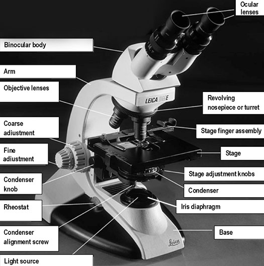
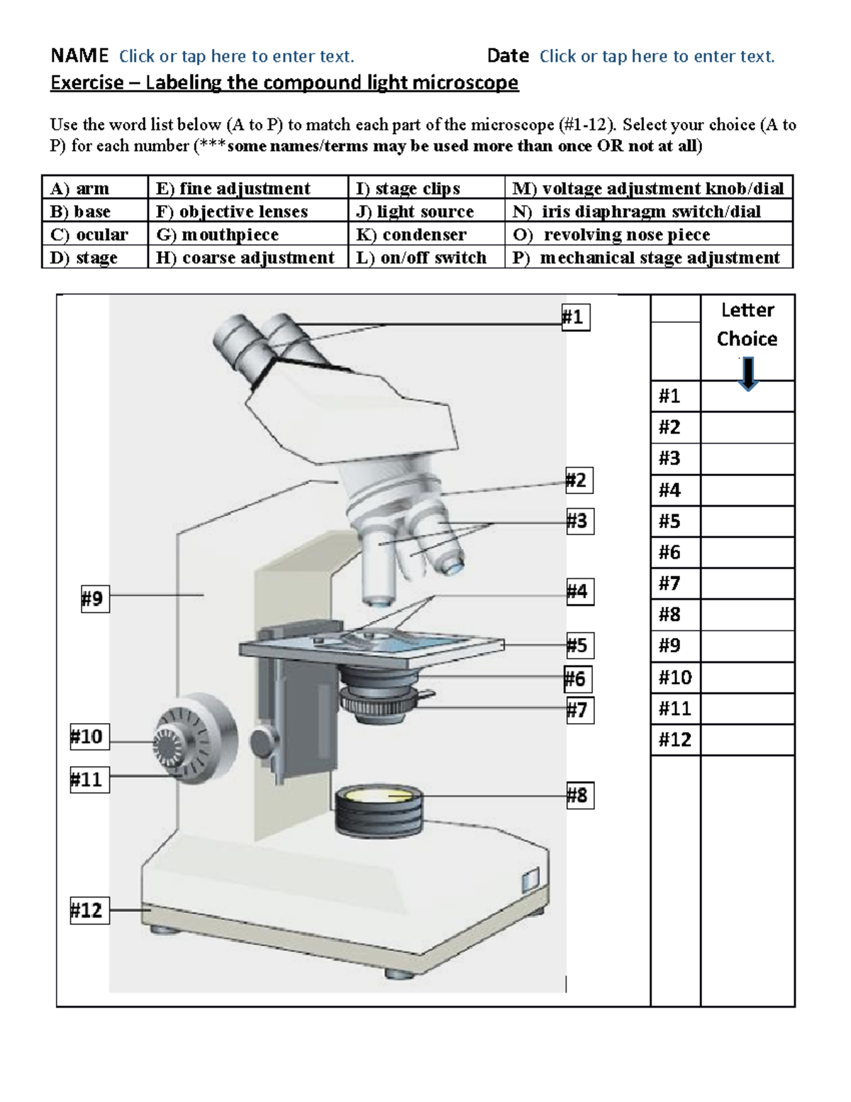
Label the Light Microscope - Labelled diagram - Wordwall Drag and drop the pins to their correct place on the image.. Eyepiece, Light Source, Base, Stage, Stage Clips, Fine Focus, Coarse Focus, Arm, Objective Lens. Compound Microscope Parts, Functions, and Labeled Diagram Compound Microscope Definitions for Labels. Eyepiece (ocular lens) with or without Pointer: The part that is looked through at the top of the compound microscope. Eyepieces typically have a magnification between 5x & 30x. Monocular or Binocular Head: Structural support that holds & connects the eyepieces to the objective lenses. Light microscope labels Flashcards | Quizlet Light microscope labels. Flashcards. Learn. Test. Match. Flashcards. Learn. Test. Match. Created by. school1329. Terms in this set (14) Ocular lens. First automatic magnification (x10) Body tube. Holds ocular lense. Revolving nose piece. Holds and allows selection of desired objective lens. Lowest power objective lens. Parts of a microscope with functions and labeled diagram Microscopic illuminator - This is the microscopes light source, located at the base. It is used instead of a mirror. It captures light from an external source of a low voltage of about 100v. Condenser - These are lenses that are used to collect and focus light from the illuminator into the specimen.
Microscope Labeling #1 Diagram | Quizlet
Microscope Parts, Function, & Labeled Diagram - slidingmotion Condenser. The condenser is to focus the light, which passes from the microscopic illuminator to the specimen. This condenser is located just below the diaphragm. This diaphragm is one of the important parts of the compound microscope which will help to get an accurate and sharp image. The condenser has a magnification power of 400X and above.

Compound Microscope Parts – Labeled Diagram and their ...
Brightfield Microscope (Compound Light Microscope)- Definition ... Brightfield Microscope Definition Brightfield Microscope is also known as the Compound Light Microscope. It is an optical microscope that uses light rays to produce a dark image against a bright background. It is the standard microscope that is used in Biology, Cellular Biology, and Microbiological Laboratory studies.

Microscope Diagram Labeled, Unlabeled and Blank | Parts of a ...
Label the microscope - Science Learning Hub All microscopes share features in common. In this interactive, you can label the different parts of a microscope. Use this with the Microscope parts activity to help students identify and label the main parts of a microscope and then describe their functions. Drag and drop the text labels onto the microscope diagram.

Compound Microscope Parts – Labeled Diagram and their ...
Light Microscope Parts Labeled - 18 images - parts of the microscope ... [Light Microscope Parts Labeled] - 18 images - optical microscopy and specimen using the transmission, microscope with labels clip art at vector clip, solved microscope parts labeling 9 label the image of a c, ,

Light Microscope - an overview | ScienceDirect Topics
Microscope Label Worksheets - K12 Workbook Displaying all worksheets related to - Microscope Label. Worksheets are The microscope parts and use, Parts of the light microscope, Label parts of the microscope, Labeling scientific tools microscope name, Parts of the microscope quiz, Use the word list to help you label the 12, Label parts of the microscope answers, Microscope lab.
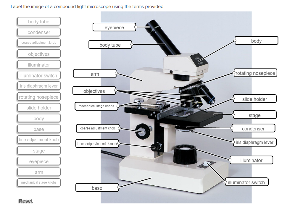
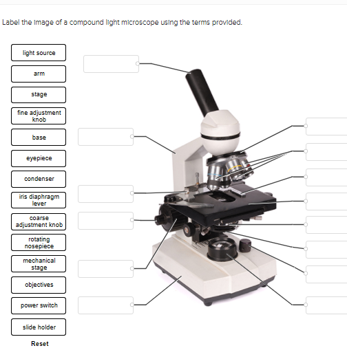
Solved Label the image of a compound light microscope using ...
Microscope Labeling - The Biology Corner 1) Start with scanning (the shortest objective) and only use the COARSE knob . Once it is focused… 2) Switch to low power (medium) and only use the COARSE knob . You may need to recenter your slide. Once it is focused.. 3) Switch to high power (long objective).
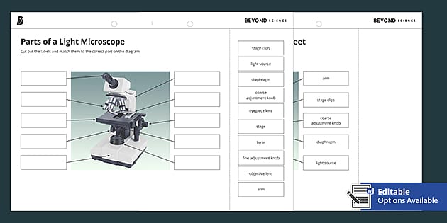
Parts of a Light Microscope Cut and Stick Worksheet - Twinkl

Compound Microscope Labeled Diagram | Quizlet

Label the microscope — Science Learning Hub

TYPES OF LIGHT MICROSCOPE: 1. SIMPLE... - Microbiologists_2 ...
Label microscope - Teaching resources
Microscope

Microscope Terms Glossary | Earth science lessons, Biology ...

Compound Microscope Parts, Diagram Definition, Application ...
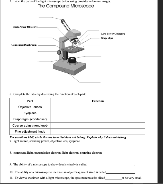
Solved 5. Label the parts of the light microscope below ...
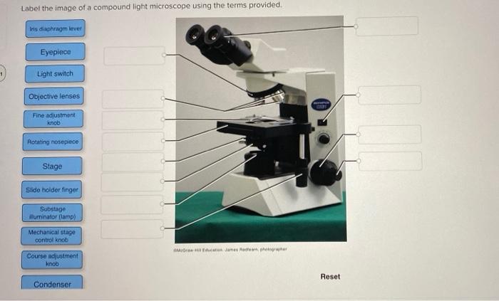
Solved Label the image of a compound light microscope using ...

Optical Microscope - an overview | ScienceDirect Topics

Microscope diagram labeled | Clipart Panda - Free Clipart Images

Labels for the light microscope for... - The Science Break ...
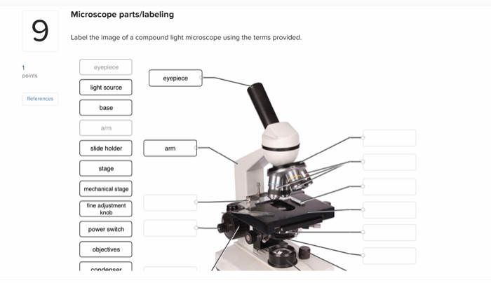
Solved Microscope parts/labeling 9 Label the image of a ...
Transmitted light microscope B3 Professional series B3-220ASC ...
Solved PLEASE HELP THANK YOU- LIGHT SOURCE WILL BE NUMBER 1 ...
Histological techniques. 6. Visualization. Light microscope ...

OMAX 40X-2500X Trinocular Biological Compound Microscope with Replaceable LED Light
9.1: Using Microscopes - Biology LibreTexts
Microscopes
Microscopes | Idaho State University

Below is a photo of a compound light microscope with labels ...
simple light microscope labeled - Clip Art Library
Label the light microscope | Teaching Resources
Solved biology 1000 lab 2.7 compound light microscope ONLY ...

Simple Microscope Definition, Magnification, Parts And Uses

Microscope Parts and Functions
Microscope - Teaching resources

Microscope, Microscope Parts, Labeled Diagram, and Functions

MICROSCOPE Labeling - Part - 3

Parts of a Compound Microscope and Their Functions
BIO 101 parts of the microscope to label - NAME Click or tap ...
Light Microscope- Definition, Principle, Types, Parts ...
Post a Comment for "39 light microscope with labels"