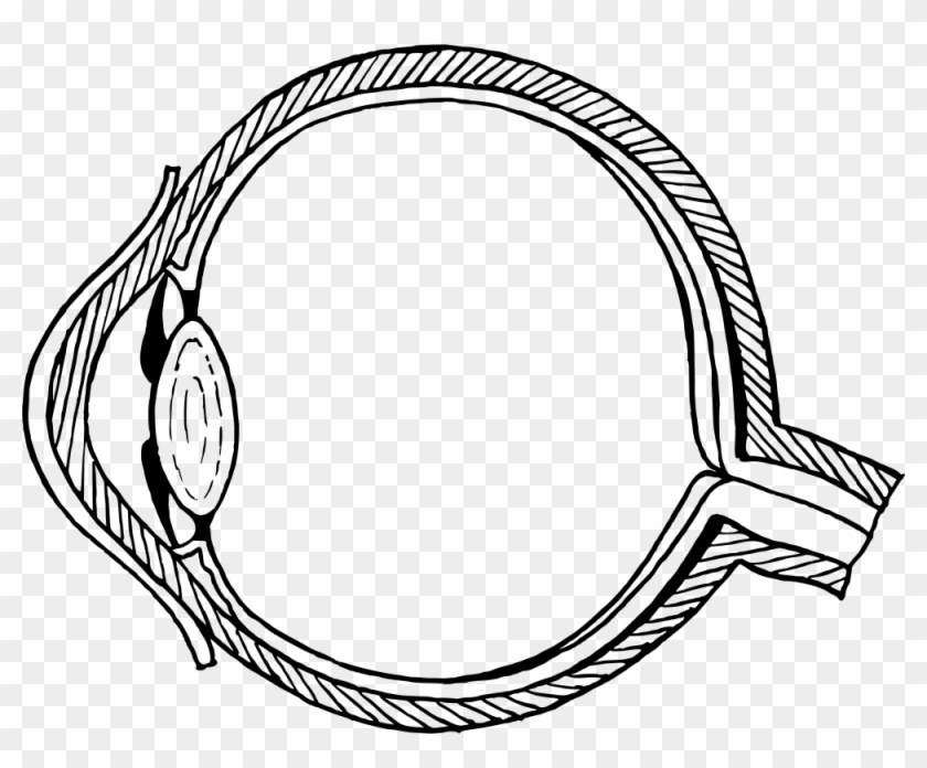
45 human eye diagram without labels
6,819 Human eye diagram Images, Stock Photos & Vectors | Shutterstock 6,819 human eye diagram stock photos, vectors, and illustrations are available royalty-free. See human eye diagram stock video clips Image type Orientation Color People Artists Sort by Popular Biology Healthcare and Medical Icons and Graphics Diseases, Viruses, and Disorders human eye anatomy 3d rendering eye medicine retina Next of 69 File:Diagram of human eye without labels.svg - Wikimedia File:Diagram of human eye without labels.svg. Size of this PNG preview of this SVG file: 410 × 430 pixels. Other resolutions: 229 × 240 pixels | 458 × 480 pixels | 732 × 768 pixels | 976 × 1,024 pixels | 1,953 × 2,048 pixels.
labelled diagram of human eye - Microsoft eye squinting human better why helps diagram science retina parts retinal without. Diagram Of Human Eye Anatomy With Label 1848847 Vector Art At Vecteezy . ... labeled eye diagram human parts vector cornea illustration similar adult. Unlabeled blank human eye diagram. Draw a well labelled diagram of human eyes.

Human eye diagram without labels
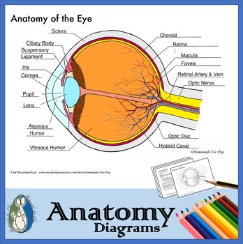
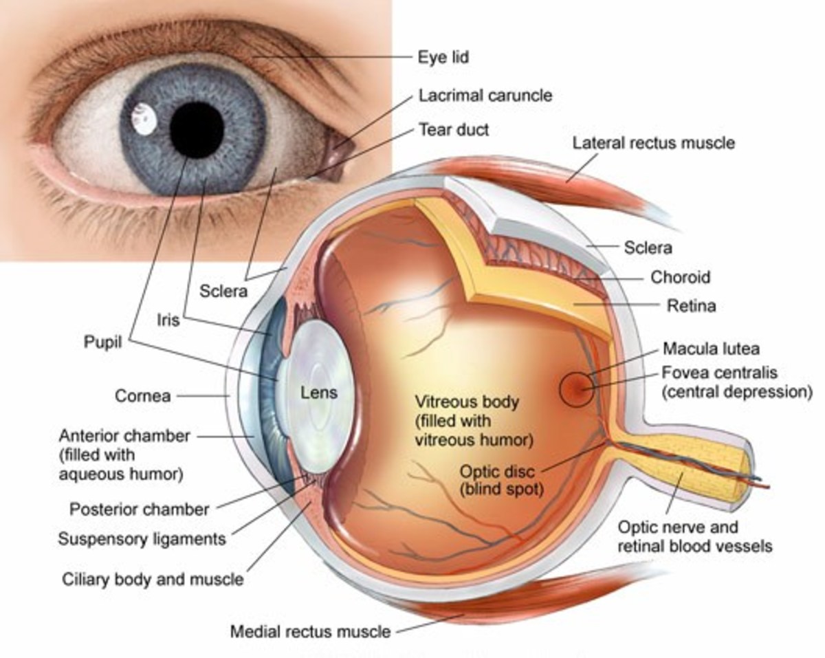
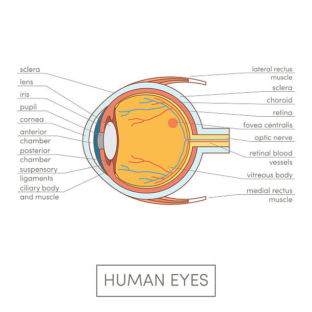
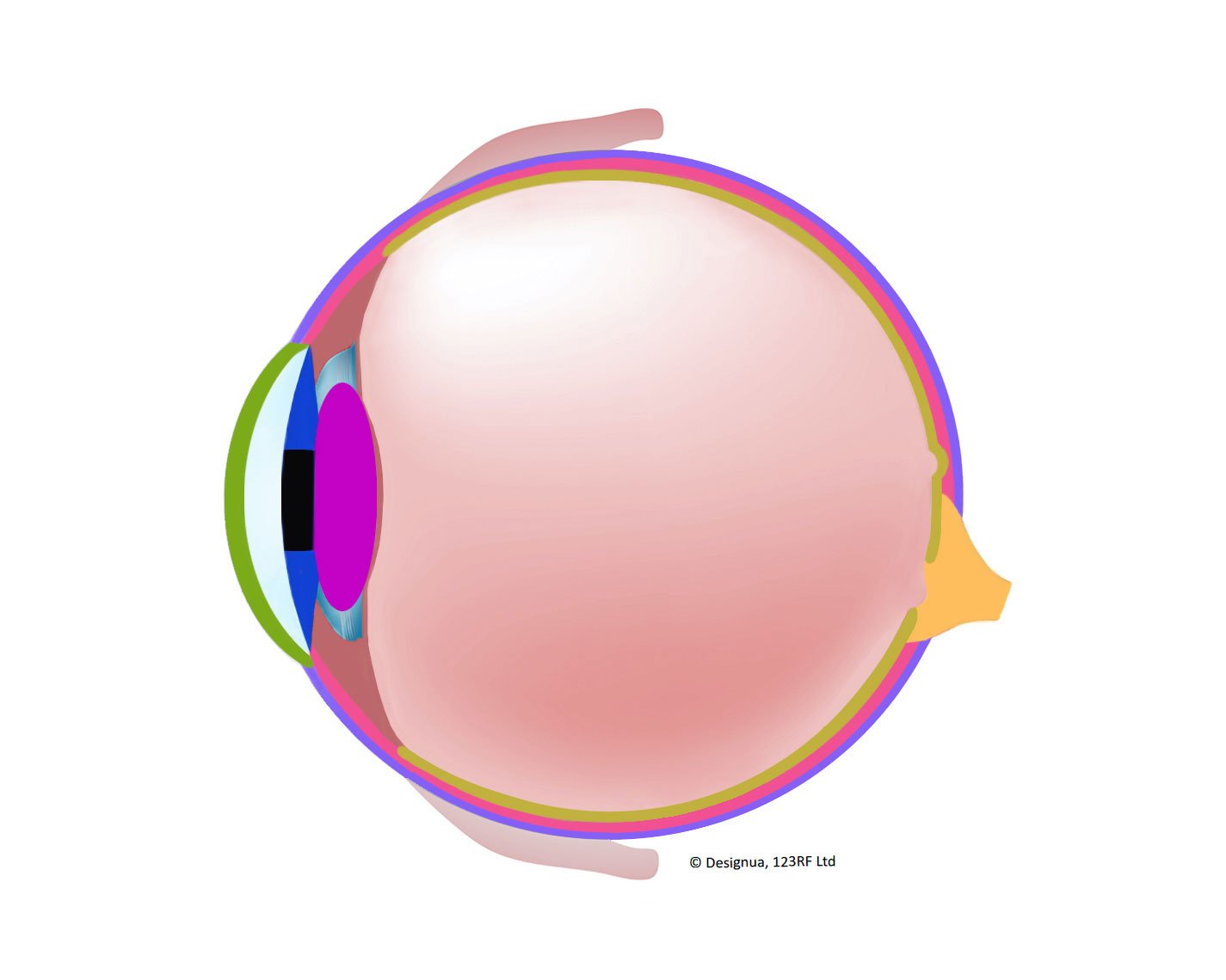
eyeball diagram labeled Eye diagram label human parts without eyes labeled structure clipart anatomy blank diagrams vision ks2 clipground pig medicinebtg figure retinal. Eye in cross section : anatomy : the eyes have it. Eye retinal artery anatomy central labels disease eyes cats diagram coloboma national institute occlusion uveal structure diseases different nih orphan The Human Eye - Diagram, Parts, Working, Function and Work of The Lens Sclera: The sclera is the protective outer layer, a strong white coating that protects the eyes (white part of the eye). Cornea: The cornea is the sclera's translucent front part. The cornea allows light to flow through and into the eye. Iris: The iris is a black muscular tissue and ring-like structure behind the cornea. The eye's colour is determined by the colour of the iris. Eye Diagram Teaching Resources | Teachers Pay Teachers The Human Eye Overview Reading Comprehension and Diagram Worksheet. by. Teaching to the Middle. 4.7. (65) $1.50. Zip. This passage briefly describes the human eye (900-1000 Lexile). 14 questions (matching and multiple choice) assess students' understanding. Students label a diagram of 6 parts of the eye.
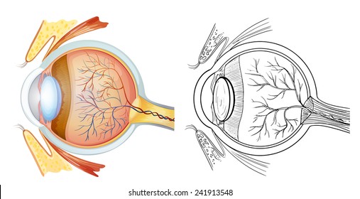
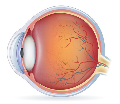
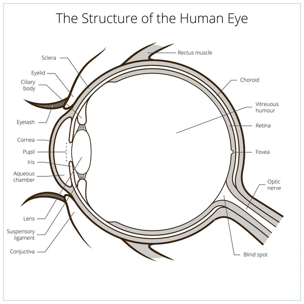
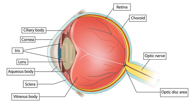
Human eye diagram without labels. What Does the Eye Look Like? - Diagram of the Eye | Harvard Eye Associates Vitreous Gel: A thick, transparent liquid that fills the center of the eye. It is mostly water and gives the eye its form and shape. Our eyes are vital for seeing the world around us. Keep them healthy by maintaining regular vision exams. Contact Harvard Eye Associates at 949-951-2020 or harvardeye.com to schedule an appointment today. File:Schematic diagram of the human eye no.svg - Wikimedia Original upload log []. This image is a derivative work of the following images: File:Schematic diagram of the human eye en.svg licensed with PD-self 2008-02-02T01:33:45Z Jakov 508x516 (54267 Bytes) suspensory ligament, arrow was wrong; 2008-01-31T16:48:11Z Jakov 508x516 (54263 Bytes) xml-Cleanup; 2007-01-25T03:10:10Z Rhcastilhos 508x516 (42056 Bytes) {{Information |Description=Schematic ... Human Ear Diagram - Bodytomy Look no further, this Bodytomy article gives you a labeled human ear diagram and also explains the functions of its different components. The human body is like a big machine, and various processes take place inside it. With the help of the various organs and tissues, it carries out some of the most marvelous tasks, that are no less than a miracle! Anatomy of the Human Eye - News-Medical.net The light passing through cornea, pupil, and lens gets focused on the retinal membrane. In addition to tissue components, retina is made up of two types of cells: rod cells and cone cells. The ...
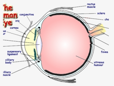
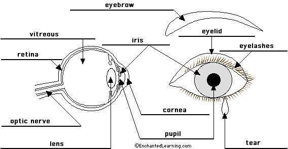
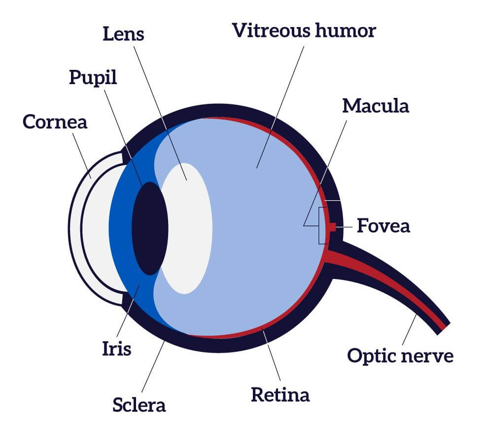
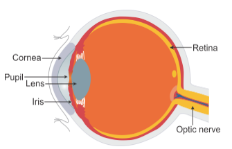
Eye diagram by Firkin | Human eye diagram, Diagram of the eye, Eye ... Eye diagram by Firkin. Find this Pin and more on free images to print by Monica Eggleton. Human Eye Diagram. Diagram Of The Eye. Eye Parts. Parts Of The Eye. Eye Anatomy. Human Body Anatomy. Free Human Body. Eye Anatomy: Parts of the Eye and How We See Behind the anterior chamber is the eye's iris (the colored part of the eye) and the dark hole in the middle called the pupil. Muscles in the iris dilate (widen) or constrict (narrow) the pupil to control the amount of light reaching the back of the eye. Directly behind the pupil sits the lens. The lens focuses light toward the back of the eye. Structure and Functions of Human Eye with labelled Diagram - BYJUS The human eye is a roughly spherical organ, responsible for perceiving visual stimuli. It is enclosed within the eye sockets in the skull and is anchored down by muscles within the sockets. Anatomically, the eye comprises two components fused into one; hence, it does not possess a perfect spherical shape. Eye Anatomy: A Closer Look At the Parts of the Eye - All About Vision In a number of ways, the human eye works much like a digital camera: Light is focused primarily by the cornea — the clear front surface of the eye, which acts like a camera lens. The iris of the eye functions like the diaphragm of a camera, controlling the amount of light reaching the back of the eye by automatically adjusting the size of the ...
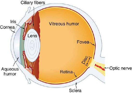
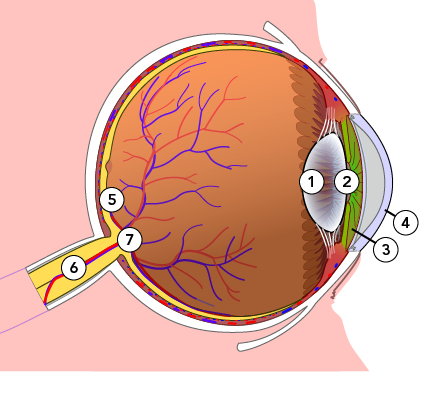
PDF Parts of the Eye - National Institutes of Health Eye Diagram Handout Author: National Eye Health Education Program of the National Eye Institute, National Institutes of Health Subject: Handout illustrating parts of the eye Keywords: parts of the eye, eye diagram, vitreous gel, iris, cornea, pupil, lens, optic nerve, macula, retina Created Date: 12/16/2011 12:39:09 PM Premium Vector | Diagram of human eye anatomy with label Download this Premium Vector about Diagram of human eye anatomy with label, and discover more than 37 Million Professional Graphic Resources on Freepik. #freepik #vector #eyeanatomy #cornea #retina The Eyes (Human Anatomy): Diagram, Optic Nerve, Iris, Cornea ... - WebMD The front part (what you see in the mirror) includes: Iris: the colored part. Cornea: a clear dome over the iris. Pupil: the black circular opening in the iris that lets light in. Sclera: the ... Human eye - Wikipedia Schematic diagram of the human eye. It shows a horizontal section through the right eye. The eye is made up of three coats, or layers, enclosing various anatomical structures. The outermost layer, known as the fibrous tunic, is composed of the cornea and sclera, which provide shape to the eye and support the deeper structures.
File:Eye Diagram without text.gif - Wikimedia Commons
Eye Anatomy: 16 Parts of the Eye & Their Functions - Vision Center The macula lutea is a yellow oval area in the retina's center (back of the eye). The center of the macula is known as the fovea. It is the section of the retina that is in charge of sharp, detailed central vision (also called visual acuity). The macula lutea has a high concentration of cones.
13,140 Eye diagram Images, Stock Photos & Vectors | Shutterstock
Eye, External Front View - resource - Imageshare - Benetech Diagram of the external view of a human eye. Design modalities for the image include braille with and without labels, print with and without labels in greyscale, color, and texture. (Source: Benetech) Metadata. Subject: Life Sciences - Science. Keywords: anatomy, diagram ...
Labelled Diagram Of Human Eye , Png Download - Label A Human ...
The Eye Diagram: What is it and why is it used? Here, the bit sequences 011, 001, 100, and 110 are superimposed over one another to obtain the example eye diagram. The eye diagram takes its name from the fact that it has the appearance of a human eye. It is created simply by superimposing successive waveforms to form a composite image. The eye diagram is used primarily to look at digital ...
25.1: The Human Eye - Physics LibreTexts
Label Parts of the Human Eye - University of Dayton Parts of the Eye. Select the correct label for each part of the eye. The image is taken from above the left eye. Click on the Score button to see how you did. Incorrect answers will be marked in red. ...
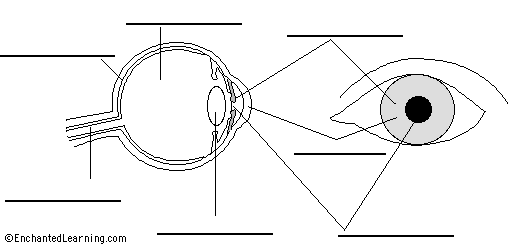
Eye Anatomy Diagram - EnchantedLearning.com
Anatomy of the eye: Quizzes and diagrams | Kenhub Take a look at the diagram of the eyeball above. Here you can see all of the main structures in this area. Spend some time reviewing the name and location of each one, then try to label the eye yourself - without peeking! - using the eye diagram (blank) below. Unlabeled diagram of the eye

Anatomy of the Human Eye
label the ear worksheet Picture Front Of The Eye Without Labels Clipart 20 Free Cliparts clipground.com eye human diagram worksheet eyeball learning layers without anatomy labels parts eyes worksheets clipart science structure grade clipground body structures 14 Best Images Of Ear Hearing Worksheets - Listening Ear Craft Template

KLB Science Interactivities - The Human Eye
PDF Eye Anatomy Handout - National Institutes of Health of light entering the eye. Lens: The lens is a clear part of the eye behind the iris that helps to focus light, or an image, on the retina. Macula: The macula is the small, sensitive area of the retina that gives central vision. It is located in the center of the retina. Optic nerve: The optic nerve is the largest sensory nerve of the eye.
/GettyImages-695204442-b9320f82932c49bcac765167b95f4af6.jpg)
Structure and Function of the Human Eye
Eye Diagram With Labels and detailed description - BYJUS A brief description of the eye along with a well-labelled diagram is given below for reference. Well-Labelled Diagram of Eye The anterior chamber of the eye is the space between the cornea and the iris and is filled with a lubricating fluid, aqueous humour. The vascular layer of the eye, known as the choroid contains the connective tissue.
Eye Diagram Images – Browse 12,983 Stock Photos, Vectors, and ...
label the eye worksheet anatomy worksheet brain eye human senses. 11 Best Images Of Parts Of The Eye Worksheet For Kids - Eye Parts . eye cow dissection diagram worksheet labeled labeling anatomy parts human label sheep teacher biologycorner lab guide blank worksheets eyes detailed. Label School Supplies Worksheet — Db-excel.com db-excel.com
Clip Art Details - Eye Diagram Without Labels - Free ...
Heart Diagram Labeled Igcse : The Human Eye Edexcel Igcse Biology ... This diagram shows the parts of the eye. 9 the diagram shows the apparatus used to demonstrate the action of amylase on starch. Veins carry deoxygenated blood and wastes from the tissues to the liver and heart. The heart's muscle is constantly active.
Eye vs. Camera | Let's Talk Science
Eye Diagram Teaching Resources | Teachers Pay Teachers The Human Eye Overview Reading Comprehension and Diagram Worksheet. by. Teaching to the Middle. 4.7. (65) $1.50. Zip. This passage briefly describes the human eye (900-1000 Lexile). 14 questions (matching and multiple choice) assess students' understanding. Students label a diagram of 6 parts of the eye.
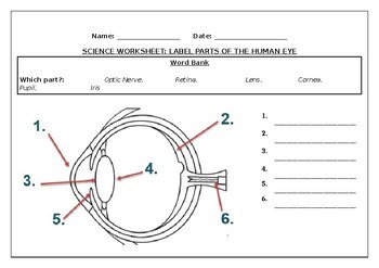
Science worksheets: Label parts of a human eye
The Human Eye - Diagram, Parts, Working, Function and Work of The Lens Sclera: The sclera is the protective outer layer, a strong white coating that protects the eyes (white part of the eye). Cornea: The cornea is the sclera's translucent front part. The cornea allows light to flow through and into the eye. Iris: The iris is a black muscular tissue and ring-like structure behind the cornea. The eye's colour is determined by the colour of the iris.

Eye Diagram Vector Art, Icons, and Graphics for Free Download
eyeball diagram labeled Eye diagram label human parts without eyes labeled structure clipart anatomy blank diagrams vision ks2 clipground pig medicinebtg figure retinal. Eye in cross section : anatomy : the eyes have it. Eye retinal artery anatomy central labels disease eyes cats diagram coloboma national institute occlusion uveal structure diseases different nih orphan

Human Eye Diagram Stock Illustration - Download Image Now ...

Cross Section of a Human Eye Diagram Black and White ...

File:Diagram of human eye without labels.svg - Wikimedia Commons
Label Eye Printout - EnchantedLearning.com
Eye diagram Vector Art Stock Images | Depositphotos

Free art print of Diagram of human eye anatomy with label

Diagram of human eye anatomy with label art print poster

Label parts of a human eye picture is there - Brainly.in
Human Eye Diagram, How The Eye Work -15 Amazing Facts of Eye
Anatomy of the Eye Diagrams for Coloring/Labeling, with ...
Anatomy and Structure of the Human Eye (With Diagrams ...

Diagram of human eye anatomy with label - Stock Illustration ...

Diagram human eye anatomy with label Royalty Free Vector

NEIBank | National Eye Institute

Human Eye Anatomy Quiz

Anatomy of the Human Eye

Structure of human eye (video) | Khan Academy

1,379 Human Eye Diagram Stock Photos, Pictures & Royalty-Free ...
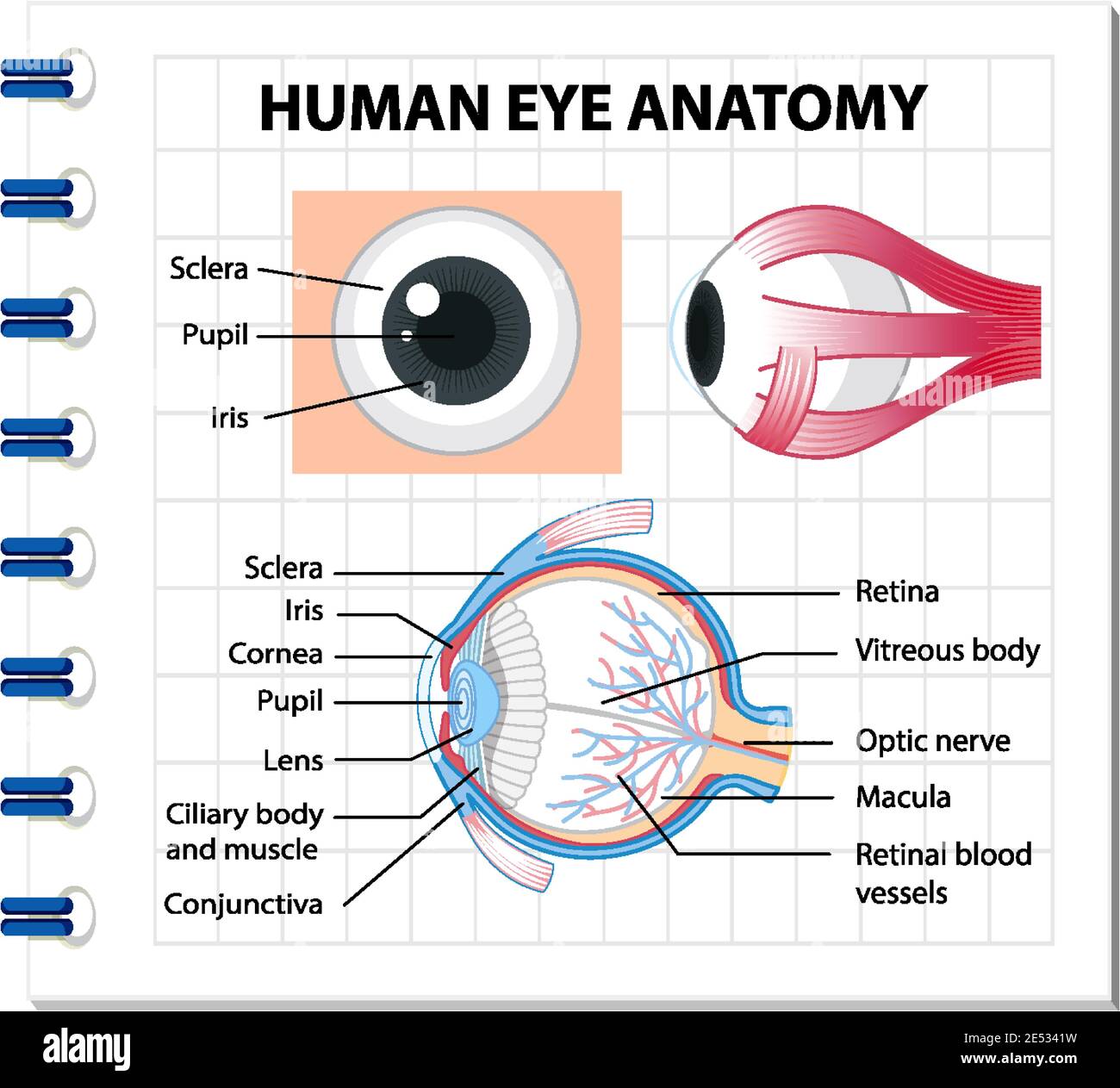
Diagram of human eye anatomy with label illustration Stock ...
4,200 Eye Diagram Stock Photos, Pictures & Royalty-Free ...
Eye Anatomy Images – Browse 47,731 Stock Photos, Vectors, and ...
Labelling the eye — Science Learning Hub

HBS 2.4 Eyes Diagram | Quizlet
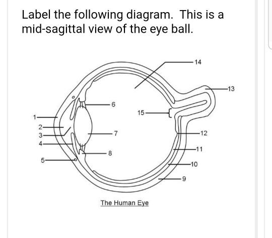
Solved Label the following diagram. This is a mid-sagittal ...
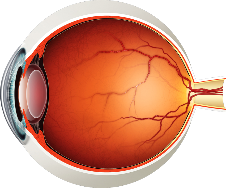
Diagram of the Eye - Lions Eye Institute
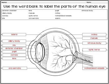
NO PREP assessment to label the HUMAN EYE

The Eye - diagram to label | Teaching Resources
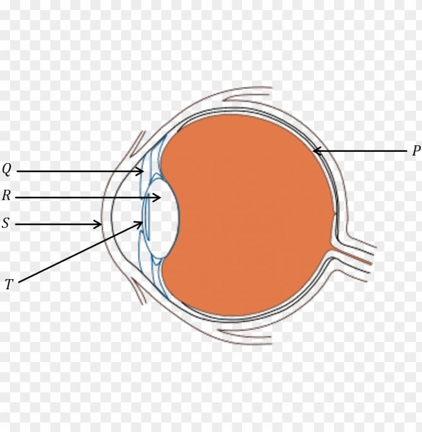
diagram of human eye - diagram of eye for class 8 PNG image ...
![Cross sectional diagram of human eye [1]. | Download ...](https://www.researchgate.net/publication/276541864/figure/fig1/AS:612895498964992@1523137082339/Cross-sectional-diagram-of-human-eye-1.png)
Cross sectional diagram of human eye [1]. | Download ...
Draw the structure of human eye and label any three parts of ...
Human Eye Coloring Page | crayola.com

Diagram of Human Eye Anatomy with Label Stock Vector ...
Post a Comment for "45 human eye diagram without labels"